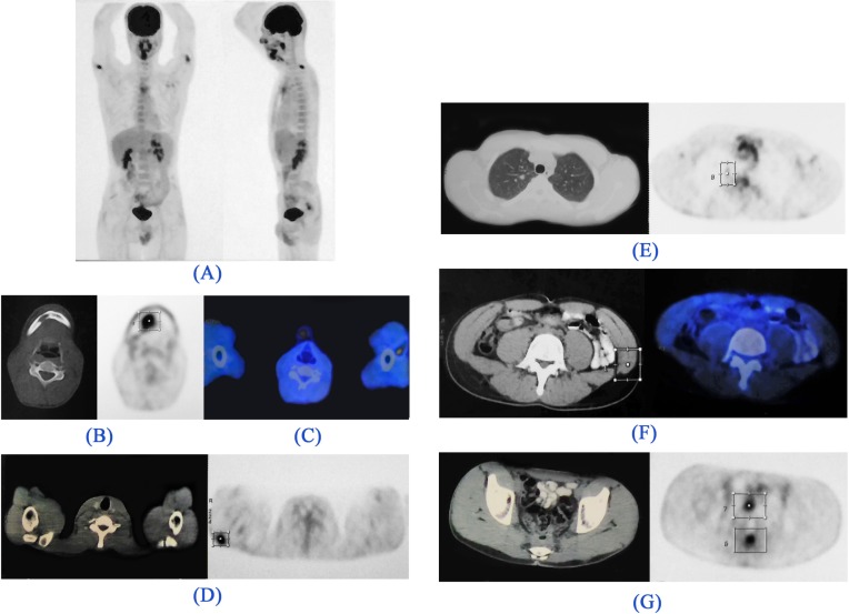
Figure 2.
18FDG-PET/CT images. (A). Maximum intensity projection (MIP) image: There are foci of metabolically active lesions in the right paramedian submandibular region, bilateral proximal upper extremities, and posterior pelvic cavity. Metabolically active lesions are demonstrated in the (B). Trans-axial CT (left) and PET (right) images: Right submandibular region (right external lingual muscles) with invasion to the adjacent mandible, (C). Trans-axial Fused image: Posterior to the left biceps muscle, (D). Trans-axial CT (left) and PET (right) images: Right deltoid muscle, (E). Trans-axial CT (left) and PET (right) images: Pulmonary nodule in the apical segment of the right lung, (F). Trans-axial CT (left) and Fused (right) images: Left external oblique abdominis muscle, (G). Trans-axial CT (left) and PET (right) images: GI tract, above the anastomotic region (which was proved to be non-malignant in subsequent colonoscopic and biopsic evaluation).

