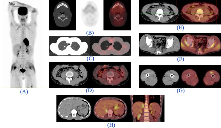
Figure 5.
18FDG-PET/CT images. (A). Maximum intensity projection (MIP): Foci of metabolically active lesions are visualized superior to the right kidney, right mid abdominal region, and the posterior pelvic cavity. In addition, diffuse metabolic activity is noted in skeletal muscles owing to physical activity in the morning of the study. Of note, due to technical issues the imaging of the lower extremities was performed separately. (B). Trans-axial CT (left), PET (middle) and Fused (right) images: Comparing with the previous study, the mandibular bone lesion is healed without evidence of remaining tumoral lesion. (C). Trans-axial CT (left) and Fused (right) images: A mild metabolically active pulmonary nodule in the apical segment of the right lung shows stable size and metabolic activity. (D). Trans-axial CT (left) and Fused (right) images: A new metabolically active lesion in the right rectus abdominis muscle. (E). Trans-axial CT (left) and Fused (right) images: A metabolically inactive lesion in the left external oblique abdominis muscle probably reveals the metabolic response to the treatment. (F). Trans-axial CT (left) and Fused (right) images: Mild metabolic activity in the GI tract, above the anastomotic region, reveals no significant change in size and metabolic activity in comparison to the previous study (which has been proven to be non-malignant in recent colonoscopic and biopsic evaluation). (G). Trans-axial CT (left) and Fused (right) images: A new metabolically active metastasis in the left quadriceps muscle. (H). Trans-axial CT (left), Fused (middle), and Coronal Fused (right) images: A new metabolically active metastatic lesion in the right adrenal gland

