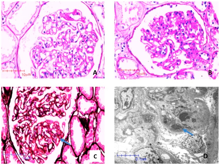
Figure 3.
Photomicrographies stained with Periodic Acid Shiff (PAS) (A) and (B) and Jones Metenamine (C), respectively (40×). The glomeruli showed segments of mesangial proliferation and endocapillar hypercellularity. Edematous endothelial cells occlude some segments (arrow). (D) Electron photomicroscopy (8000×). Subendothelial electron dense deposits (immune complexes) are indicated by arrows.

