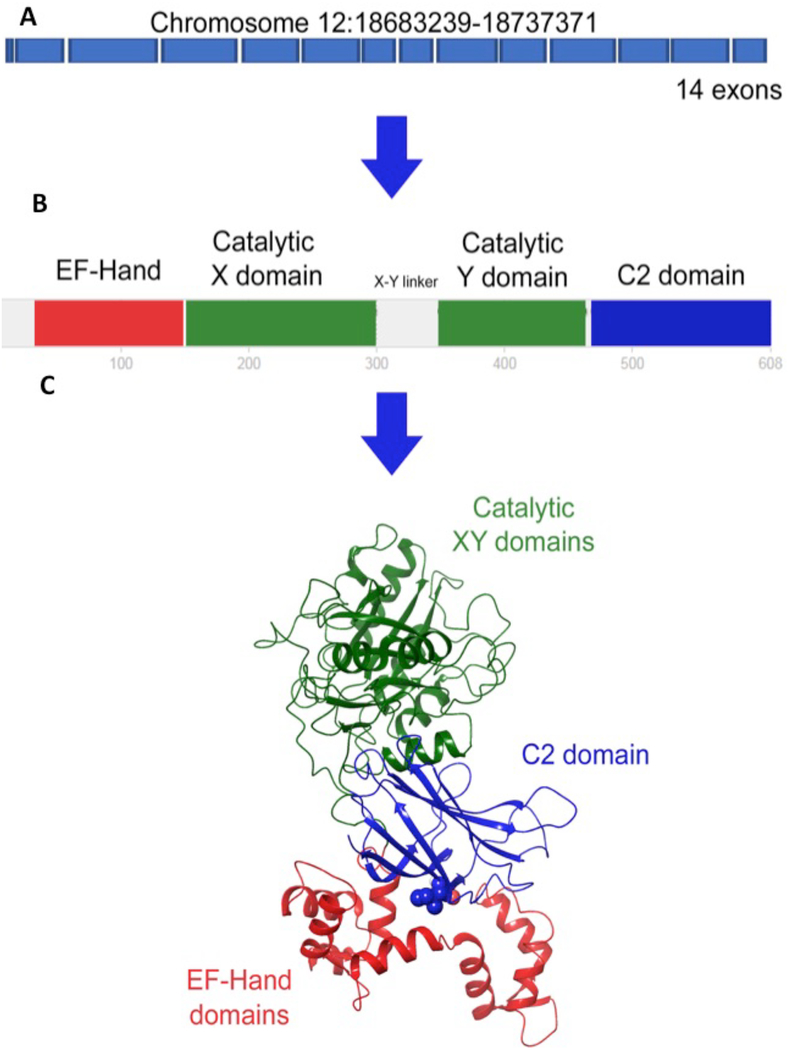
Figure 3. Organization of the different domains of PLCζ.
A. The PLCZ1 gene contains 15 exons. The coding sequence starts at exon 2 and finishes at exon 15. B. PLCζ has three different domains, which are the EF-hands, the catalytic domain, and the C2 domain from the N terminus to the C terminus, respectively. The catalytic domain is subdivided into X and Y catalytic regions joined by the XY linker. The X and Y domains act together on the target as two mandibles of a jaw. C. The 3D structure of hPLCζ was modeled from the crystallographic structure of rPLCδ1. The model shows the close spatial interaction of the EF-hand and the C2 domains. The human I489F mutation is located at the EF-hands-C2 domain interface. Adapted from Escoffier et al, 2016.

