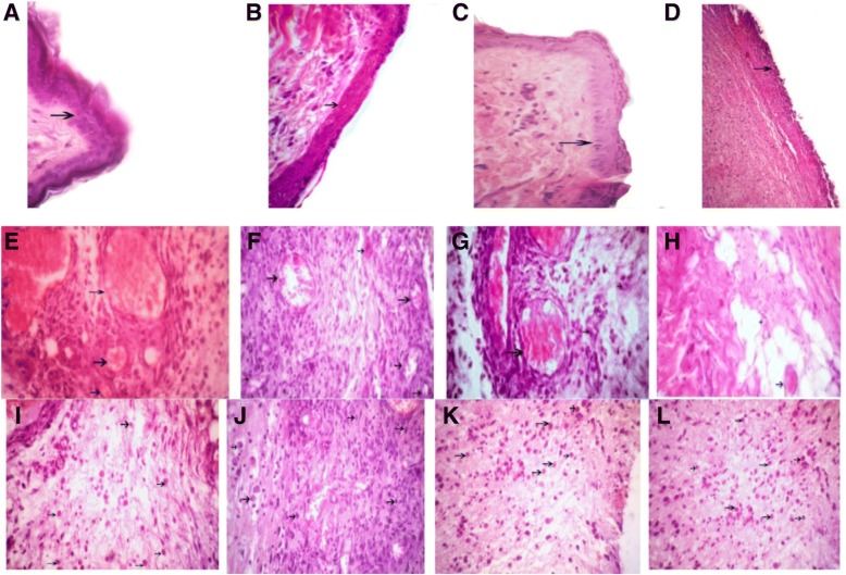
Fig. 5.
Arrows show the wound epidermis regeneration (a–d), vascular formation (e–h), and granulation (i–l) on the third day. a, e, i Satureja khuzistanica-treated group. b, f, j Hydrogel alginate-treated group. c, g, k Satureja khuzistanica encapsulated in hydrogel alginate-treated group. d, h, l Control group. Hematoxylin and eosin stain (× 400). Arrows show the re-epithelialization with mild hyperplasia of the epidermis and moderate hyperkeratosis (a–d) and immature granulation tissue (i–l) with emerging blood vessels (angiogenesis) shown in black arrows and fibroblasts and numerous macrophages

