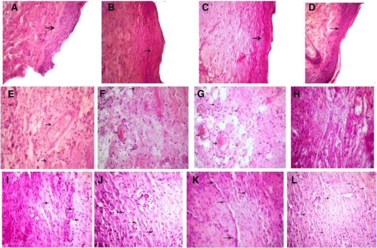
Fig. 6.
Arrows show the wound epidermis regeneration (a–d), vascular formation (e–h), and granulation (i–l) on the seventh day. a, e, i Satureja khuzistanica-treated group. b, f, j Hydrogel alginate-treated group. c, g, k Satureja khuzistanica encapsulated in hydrogel alginate-treated group. d, h, l Control group. Arrows show the re-epithelialization with mild hyperplasia of the epidermis and moderate hyperkeratosis (a–d) and immature granulation tissue (i–l) with emerging blood vessels (angiogenesis) shown in black arrows and fibroblasts and numerous macrophages. Hematoxylin and eosin stain (× 400)

