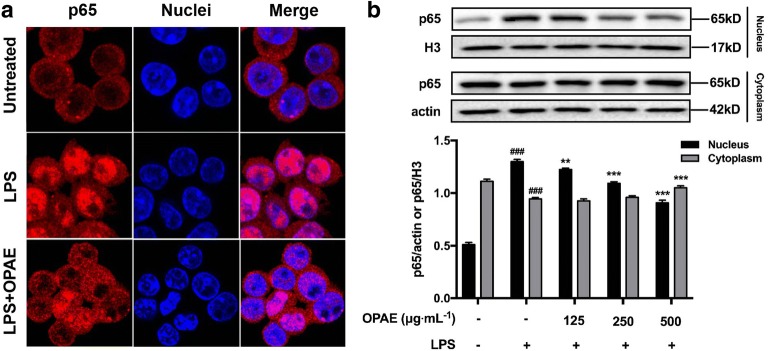
Fig. 6.
OPAE reduced NF-κB translocation into nucleus in LPS-treated RAW264.7 macrophages. Cells were pre-treated with OPAE at different concentrations (125, 250, and 500 mg mL−1) for 4 h and then stimulated with LPS (1 µg mL−1) for another 30 min. a The transcription factor, p65, was stained with primary p65 antibody followed by Alexa Fluor 568 dye conjugated secondary antibody (red fluorescence) and Hoechst 33342 dye (blue fluorescence), sequentially. b Nuclear and cytosolic proteins were subjected to Western blot analysis with the indicated anti-bodies. β-actin and histon H3 were used as internal controls for the cytosolic and nuclear fractions, respectively. Data are expressed as mean ± S.D. ###p <0.001 vs. Ctrl. group, **p <0.01, ***p <0.001 vs. LPS group

