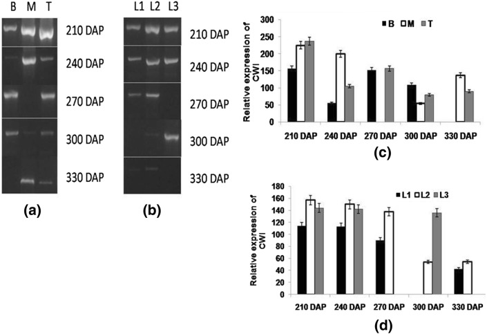
Fig. 5.
Semi-quantitative reverse transcriptase PCR (end-point) analysis of CWI a stalk samples viz. bottom (B) middle (M) top (T) b leaf samples viz. source (L1), LTM(L2), sink (L3) in CoJ64 at 210, 240, 270, 300 and 330 DAP. Intensities of amplified bands depicting variations in expression of CWI gene in c stalk and d leaf samples

