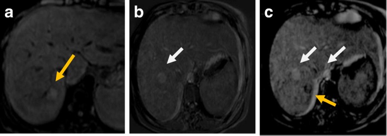
Fig. 2.

MRI after RF. a Pre-contrast T1-WI fat-suppressed image shows the ablated HFL at subsegment VII (yellow arrow) with its bright signal due to coagulative necrosis. b Subtracted arterial phase at a higher level shows slice mis-registration between the pre-contrast and post-contrast image and mirror image of the aorta (white arrow). c Subtracted arterial phase at the HFL shows slice mis-registration between the pre-contrast and post-contrast image and mirror image of the aorta (white arrow) with eccentric faint enhancement at the lesion (not consistent with the residual viable tumor). Final diagnosis is LR-TR nonevaluable due to image degradation
