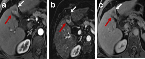
Fig. 6.

Hepatic focal lesion after RFA. a Enhanced T1-WI in the arterial phase shows early arterial peripheral nodular enhancement (red arrow). b Subtracted T1-WI arterial phase shows true enhancement of the nodule (red arrow). c Enhanced T1-WI in the delayed phase shows washout with capsule enhancement (red arrow). Overall post-treatment assessment is LR-TR viable
