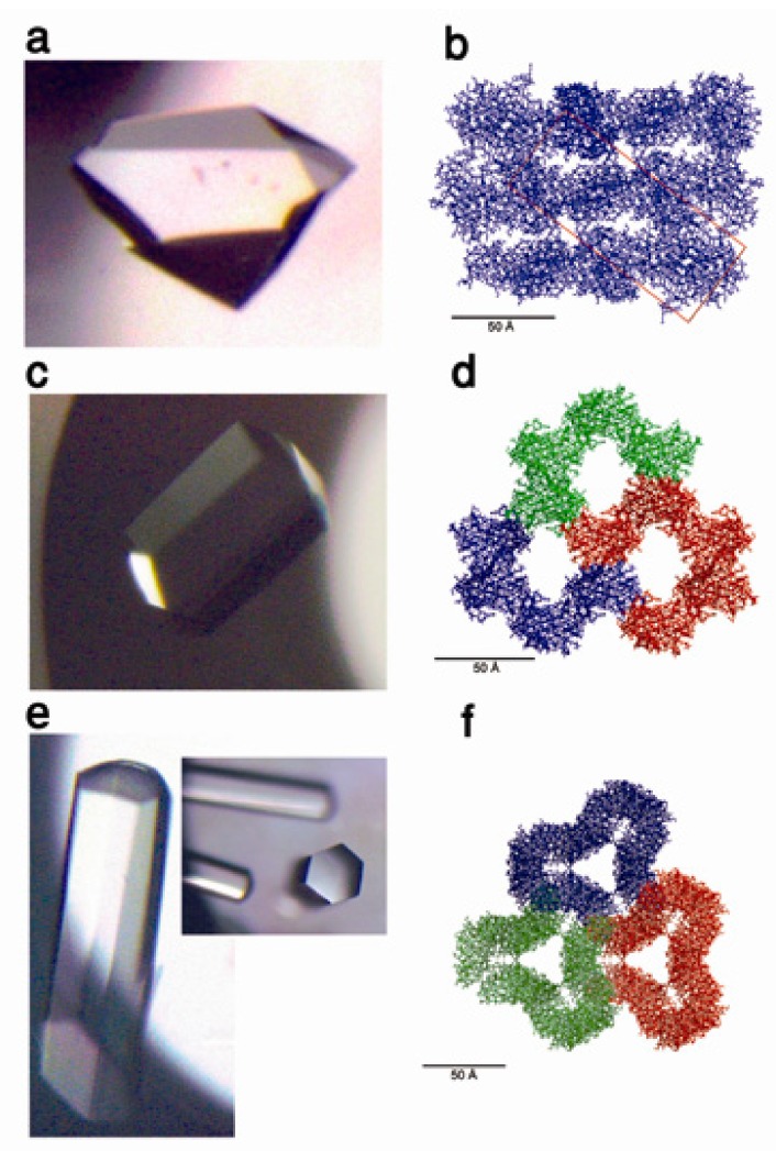
Figure 5.
(a) Optical micrograph of a parent cohesin 1 (Coh1) crystal and (b) ribbon representation of its crystal packing (cross-section in bc plane). (c) Optical micrograph of a K42W mutant protein crystal and (d) ribbon representation of its crystal packing (cross-section in bc plane). (e) Optical micrograph of a K117W-K119W double mutant protein crystal and (f) ribbon representation of its crystal packing (cross-section in bc plane). Reprinted with permission from [83], Copyright 2010, AIP Publishing LLC.

