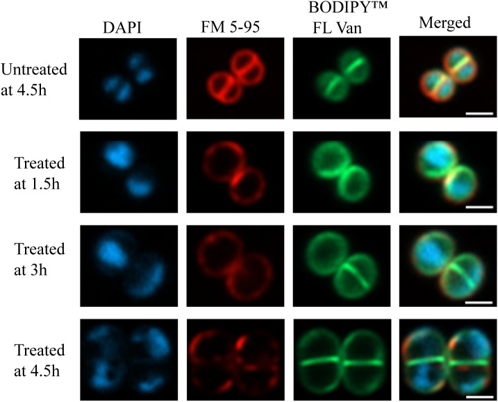
FIGURE 5.
Super-resolution fluorescence microscopy of PPAP 23 treated S. aureus. Fluorescent images of untreated S. aureus HG001 at 4.5 h and 2× MIC PPAP 23 treated cells at 1.5, 3, and 4.5 h. Cells were labeled with (left to right) DAPI (nucleoid), FM 5-95 (membrane) and BODIPYTM FL vancomycin (cell wall) and merged. The scale bar is 1 μm.

