Figure 5.
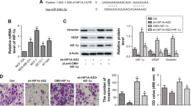
sh-HIF1A-AS2 inhibited cell motility via regulating HIF-1α/VEGF signaling pathway in MCF-7 cells.
Notes: MCF-10A and BC cells lines (MDA-MB-231, MCF-7, ZR-75–1, and BT-549) were incubated under 1% O2 for 48 hours. (A) The target of miR-548c-3p on the HIF1A 3′UTR. (B) The expression level of HIF-1α was detected by RT-qPCR (*P<0.05 vs control; **P<0.01 vs control). (C) Expression of EMT marker proteins, VEGF, and HIF-1α was measured by Western blot in MCF-7 cells transfected with sh-HIF1A-AS2 or pLenti-CMV-HIF-1α and in combination with pLenti-CMV-HIF-1α (*P<0.05 vs control, #P<0.05 vs CMV-HIF-1α group). (D) Invasion ability of MCF-7 cells transfected with sh-HIF1A-AS2 or pLenti-CMV-HIF-1α and in combination with pLenti-CMV-HIF-1α. Invasive cells were stained with crystal violet solution, and the quantification of invasive cells is shown (*P<0.05 vs control, #P<0.05 vs CMV-HIF-1α group). Scale bar =20 µm. (E) Proliferation ability was measured by CCK-8 assays in MCF-7 cells transfected with sh-HIF1A-AS2 or pLenti-CMV-HIF-1α and in combination with pLenti-CMV-HIF-1α (*P<0.05 vs control, #P<0.05 vs CMV-HIF-1α group).
Abbreviations: BC, breast cancer; CCK-8, Cell Counting Kit-8; Ctrl, control; EMT, epithelial -mesenchymal transition; HIF1A-AS2, hypoxia-inducible factor-1 alpha antisense RNA-2; RT-qPCR, quantitative real-time PCR; HIF-1α, hypoxia-inducible factor-1 alpha; UTR, untranslated region; VEGF, vascular endothelial growth factor.

