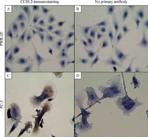
Fig.1.

Immunocytochemical staining reveals overexpression of CCRL2 in PC3 cells. Cells were incubated with mouse/IgG, monoclonal,anti-human-CCRL2 antibody followed by the respective secondary antibody. The detection was performed with EXPOSE Mouse and Rabbit Specific HRP/AEC Detection IHC system. PWR-1E cells with negative cytoplasmic staining (A). PC3 cells with strong cytoplasmic staining (C). Cells stained with no primary antibody served as negative controls for each cell line: PWR-1E (B); PC3 (D); Magnification: 400×; Brown color: AEC; Blue color: hematoxylin counterstain.
