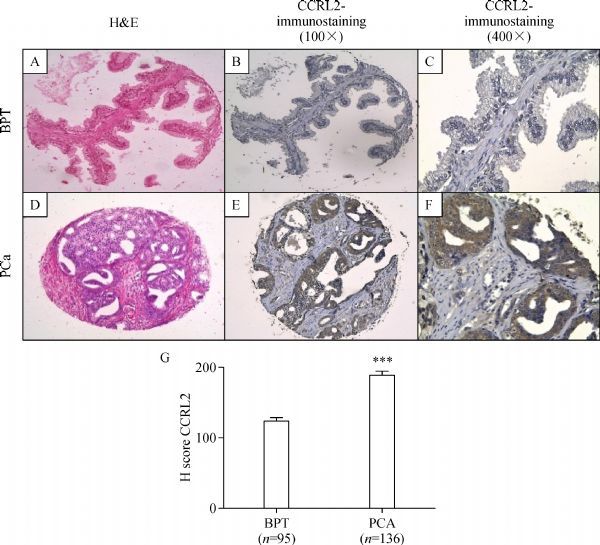
Fig.2.

Immunohistochemical staining of CCRL2 in BPT and PCa in a human prostate tissue microarray. BPT in a prostatectomy tissue spot with H&E staining 100×(A), immunohistochemical staining of CCRL2 showing absent to mild staining of CCRL2 in the epithelial compartment (B and C). Prostate cancer (PCa) tissue in a prostatectomy tissue spot with H&E staining 100×(D), immunohistochemical staining of CCRL2 showing strong staining in tumor compartment (E and F). H-score of immunohistochemical (IHC) staining in prostate tissues (G). Hscores were calculated as described in Materials and methods. *** P < 0.0001, Mann Whitney test.
