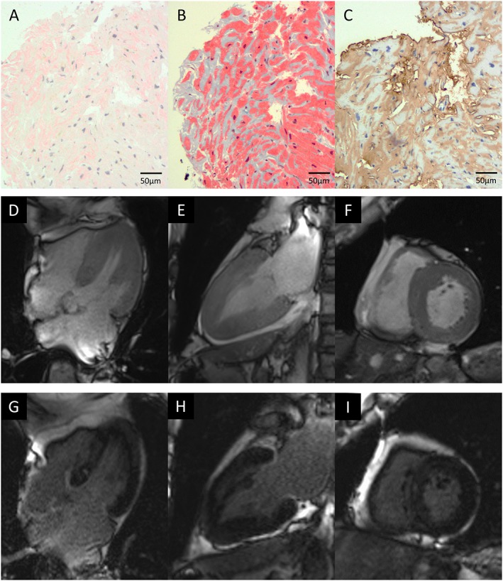
Figure 1.

Endomyocardial biopsy and cardiac magnetic resonance imaging. (A) Congo red staining and (B) Masson's trichrome staining revealed diffuse amyloid deposits in the myocardial interstitium, which were (C) immunopositive for transthyretin. Cardiac magnetic resonance imaging prior to tafamidis showed diffuse left ventricular hypertrophy by cine imaging ((D) four‐chamber view; (E) two‐chamber view; (F) short‐axis view) and diffuse late gadolinium enhancement of the left ventricle ((G) four‐chamber view; (H) two‐chamber view; (I) short‐axis view).
