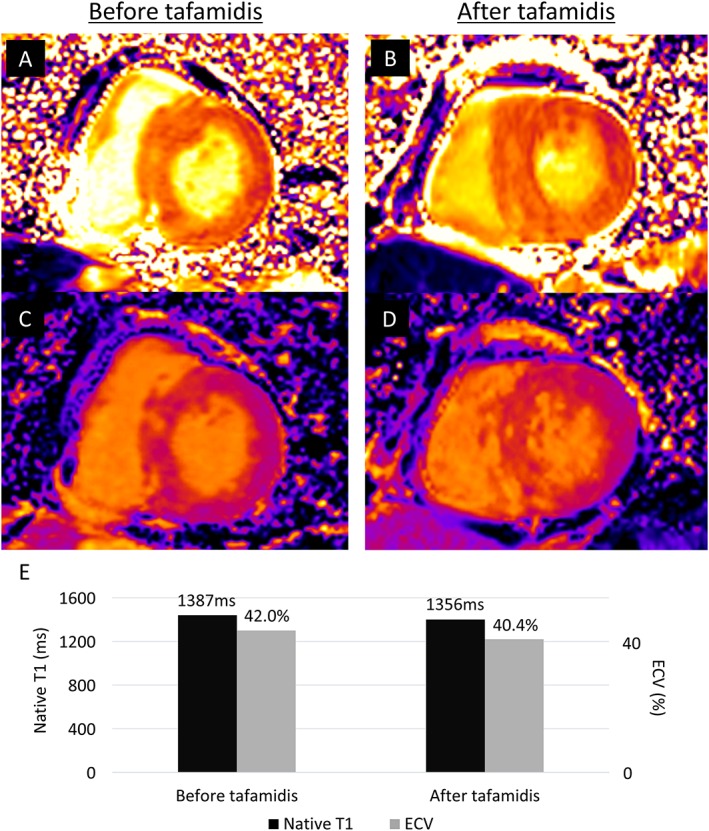
Figure 2.

Native T1 and extracellular volume (ECV) before and after 12 months of tafamidis treatment. Three tesla cardiac magnetic resonance imaging showing native T1 mapping (A) before and (B) after tafamidis. There was no obvious worsening of native T1 value (1387 to 1356 ms). ECV (C) before and (D) after tafamidis also showed no significant worsening (42.0 to 40.4%). The serial changes are summarized in (E).
