Figure 3.
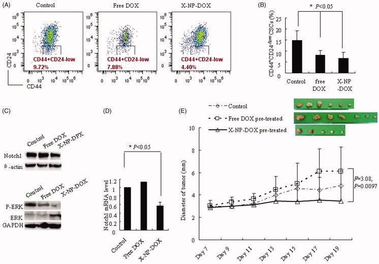
Low-dose X-NP-DOX inhibits CSCs of MCF-7 breast cancer cells. The CSC percentages assay, western blotting analysis, quantitative real-time PCR analysis, and nude mouse orthotopic breast cancer model were to further investigate the potential mechanisms of low-dose X-NP-DOX CSC inhibition. (A) The flow cytometry analysis of CSC population in MCF-7 cells with or without free DOX or X-NP-DOX pretreatment. (B) The percentages of CSCs with low-dose X-NP-DOX or free DOX were significantly lower than that of PBS-treated cells (p < .05). (C) Western blotting analysis revealed that low-dose X-NP-DOX strongly inhibited the expression of Notch1 and phosphor-Erk1/2 compared with free DOX or control. (D) Quantitative real-time PCR analysis showed that Notch1 gene expression was suppressed by low-dose X-NP-DOX. E: In brief, 100% mice were induced tumor injected by MCF-7 cells pretreated by DOX (7/7), whereas the percentages of detected tumor-bearing mice in control or X-NP-DOX-pretreated group were 85.7% (6/7) and 71.4% (5/7), respectively. The results showed that tumor growth was inhibited by X-NP-DOX pretreatment, whereas continuous tumor growth was observed in mice treated with free DOX (F = 3.08, p = .0097).

