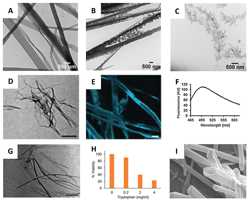
Figure 4.
Tyr and Trp self-assembly. (A–C) TEM micrographs of Tyr assemblies at ~ 4 mM at various time points: (A) 6 days, (B) 14 days, (C) 22 days.[14a] (D–F) Amyloid nature of Tyr fibers. (D) TEM image (scale bar 500 nm), (E) ThT microscopy (scale bar 20 μm) and (F) fluorescence spectrum of Tyr fibers.[14b] (G) TEM image of Trp fibers (scale bar 500 nm).[14c] (H) Cytotoxicity of Trp fibers towards SH-SY5Y cells.[14c] (I) FESEM images of Trp nanotubes (scale bar 200 nm).[35] Figures reproduced with permission from respective references.

