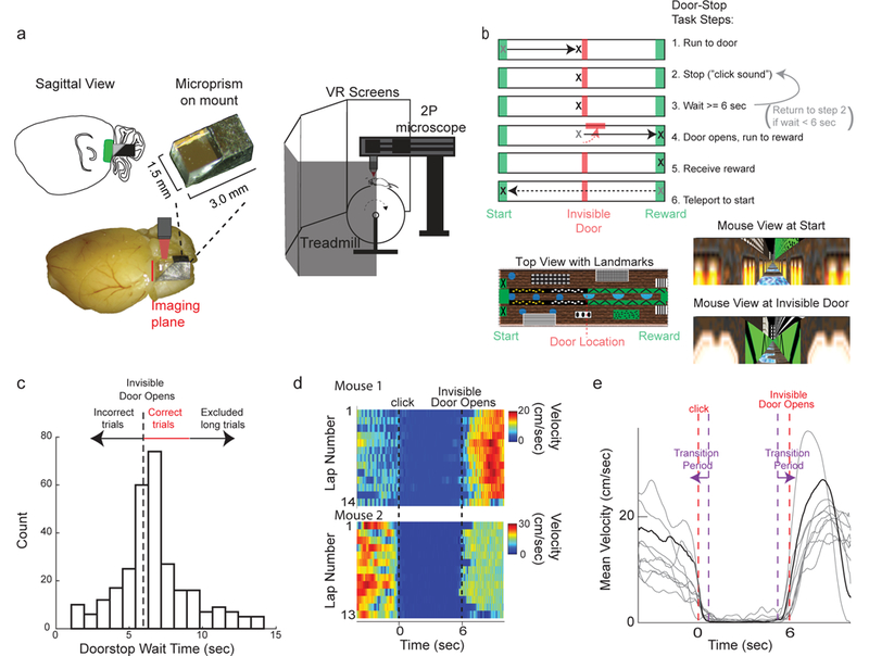
Figure 1:

Head-fixed cellular resolution functional imaging during mouse navigation in a virtual Door Stop Task. a. MEC was imaged through a microprism (left) with two-photon microscopy in head-fixed mice navigating in a virtual “Door Stop” task (right). b. Virtual Door Stop task. c. Histogram of wait times at the invisible door from all mice (n=7) trained on 6-second Door Stop task. d. Mouse locomotion velocity leading into (time < 0 sec), during (0 sec < time < 6 sec) and after 6 second Door Stop wait interval for all correct trials from 2 different single sessions in 2 mice (top and bottom). e. Mean velocity leading into (time < 0 sec), during (0 sec < time < 6 sec) and after 6 second Door Stop wait interval across all correct trials of an example single session (grey). Mean velocity across all trials for example session shown in black. Purple dashed lines and arrows indicate Transition Period.
