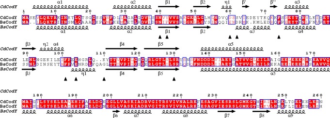
Fig 4. Alignment of the sequences of CodY from C. difficile (Cd) and B. subtilis (Bs).
The protein secondary structures for the full-length CdCodY and BsCodY are depicted above and below the sequences, respectively. Sequence identities are indicated by red shading. Residues that form prominent interactions with the effector molecule (isoleucine) in C. difficile CodY are labeled with black arrowheads.

