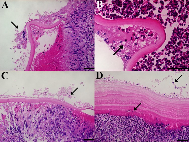
Fig 5. Cells with big nuclei within the germinal layer of both infertile and small hydatid cysts.
Histological sections of cyst wall samples with the germinal, laminated and adventitial layers in succession. The arrow points at cells with bigger nuclei than parasite cells, either inside the laminated layer or within the germinal layer. Stained with H&E. Size bar: 50 μm.

