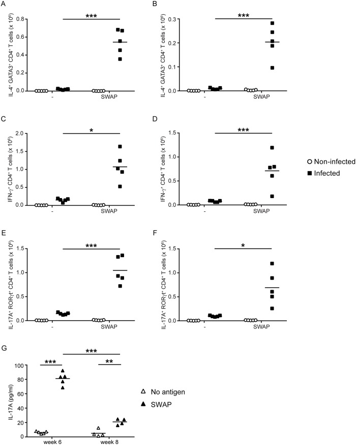
Fig 6. TH2, TH1 and TH17 cells induced by acute schistosome infection proliferate and produce cytokine in response to schistosome worm antigens.
At 6 weeks post infection, cells from the spleens (A, C, E) and mesenteric lymph nodes (B, D, F) of S. mansoni-infected mice were cultured in vitro in the absence (-) or presence of S. mansoni worm antigens (SWAP). Spleen and mesenteric lymph node cells from non-infected mice were included as controls and cultured under identical conditions. After three days in culture, intracellular cytokine/transcription factor staining and flow cytometry were used to determine the number of IL4+ GATA3+ CD4+ T cells (A, B), IFN-γ+ CD4+ T cells (C, D), and IL-17A+ RORγt+ CD4+ T cells (E, F) in the cultures. G, spleen cells from mice that were infected with S. mansoni for 6 and 8 weeks were cultured in vitro either in the presence or absence of S. mansoni worm antigens (SWAP). After three days of culture, the concentration of IL-17A in the culture supernatant was determined by ELISA. *, P < 0.05; **, P < 0.01; ***, P < 0.001.

