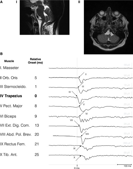
Figure 1.

(A) Saggital (1) and transversal (2) T2‐weighted MR images showing elongation of the left vertebral artery with left‐sided compression of the lateral medulla oblongata. (B) Recruitment sequence of 10‐channel polymyography. Right‐sided muscles are specified on the left. Arrows with corresponding (latin) muscle number represent the onset of muscle recruitments. Corresponding latencies (in ms), relative to the trapezius muscle (bold), are depicted on the right.
