Figure 1.

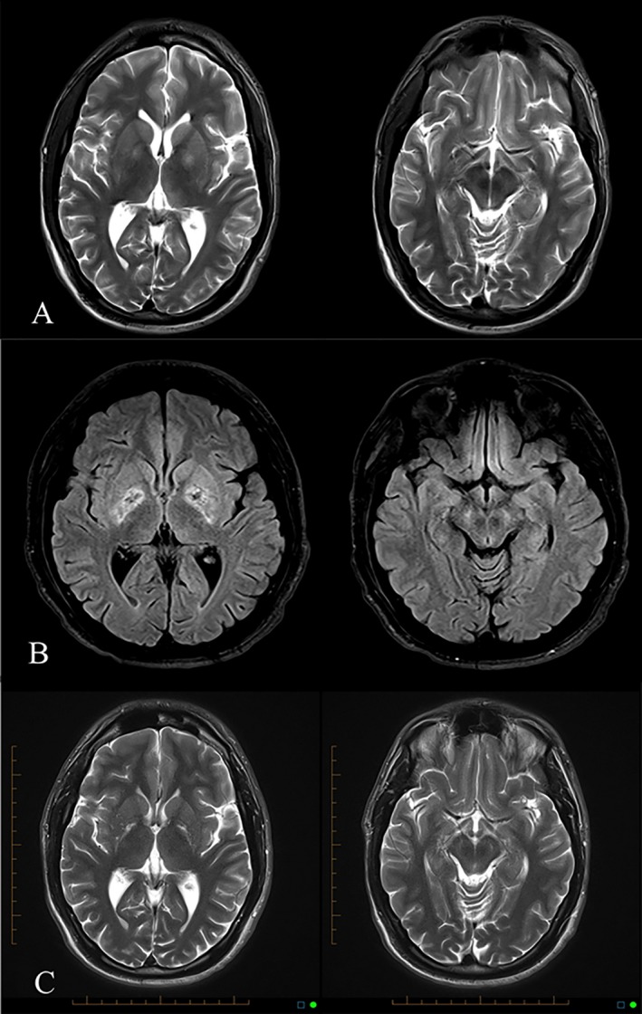
(A) T2‐weighted MRI after overdose showing GP hyperintensity and normal SN. (B) FLAIR MRI 1 month after overdose showing mixed signal in GP and new high signal in SN. (C) T2‐weighted MRI 3 years after overdose showing gliosis of GP and SN.

(A) T2‐weighted MRI after overdose showing GP hyperintensity and normal SN. (B) FLAIR MRI 1 month after overdose showing mixed signal in GP and new high signal in SN. (C) T2‐weighted MRI 3 years after overdose showing gliosis of GP and SN.