
Figure 1.
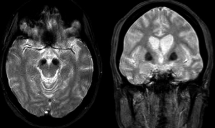
Brain MRI showing iron accumulation. The marked hypointensity in the basal ganglia (especially in the internal globus pallidus) and substantia nigra suggests iron deposition.
Official websites use .gov
A
.gov website belongs to an official
government organization in the United States.
Secure .gov websites use HTTPS
A lock (
) or https:// means you've safely
connected to the .gov website. Share sensitive
information only on official, secure websites.
Brain MRI showing iron accumulation. The marked hypointensity in the basal ganglia (especially in the internal globus pallidus) and substantia nigra suggests iron deposition.