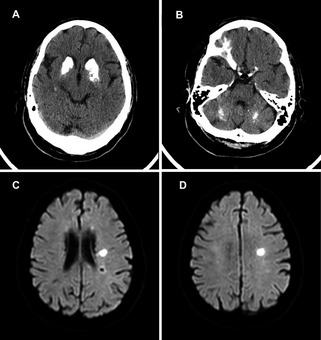
Figure 1.

(A and B) Patient's brain CT scan at age 65, showing extensive bilateral calcification of basal ganglia and dentate nuclei in the cerebellum. (C and D) Patient's brain MRI on admission, demonstrating a left frontal lacunar stroke in the periventricular white matter.
