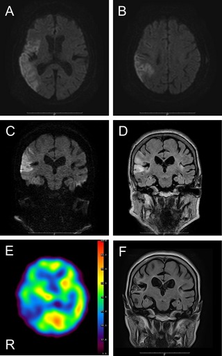
Figure 1.

(A and B) Axial diffusion‐weighted images DWIs demonstrated acute infarction in the right temporal‐parietal lobe. (C) Coronal DWI and (D) fluid‐attenuated inversion recovery verified no lesions in the basal ganglia. (E) SPECT showed hyperperfusion in the right basal ganglia. (F) Follow‐up MRIs detected no lesions in the basal ganglia.
