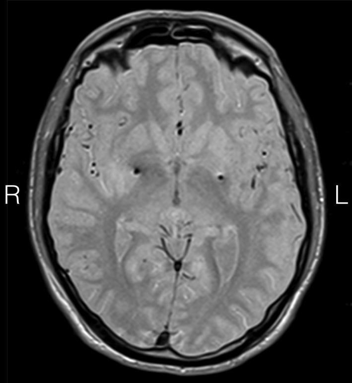
Figure 1.

Stereotactic axial 2‐mm‐thick proton density MR scan at the level of anterior/posterior commissure showing electrode (model 3389; Medtronic, Inc., Minneapolis, MN) artifacts in the posterior GPi. Active contact coordinates measured from the mid‐commissural point were: left GPi (contact 1): x = −22.15, y = 2.63, z = −1.76; right GPi (contact 10): x = 20.86, y = 5.51, z = −1.02.
