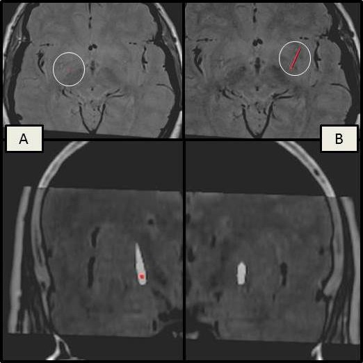
Figure 1.

Images, using FrameLink, of the preoperative MRI fused with postoperative stereotactic CT for electrode position evaluation, showing a correct positioning of the left electrode (A) within the posteroventral lateral GPi and a lateral trajectory of the right electrode ending within the GPe (B).
