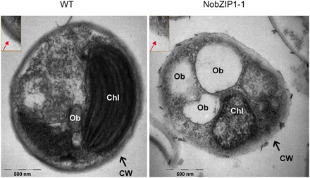
Fig. 2. Ultrastructural analyses of NobZIP1-engineered cells.

WT cells were encapsulated by rigid cell wall (left), and the enlarged image of cell wall was given in the box; NobZIP1-overexpressing cells exhibited loosen cell wall and highly enriched in oil bodies (middle). Enlarged image of cell wall was given in the box and the alteration in the cell wall structure was indicated by arrow marks. CW, cell wall; Chl, chloroplast; Ob, oil bodies. Scale bars, 500 nm.
