
Figure 1: Circadian circuits in the fly brain.
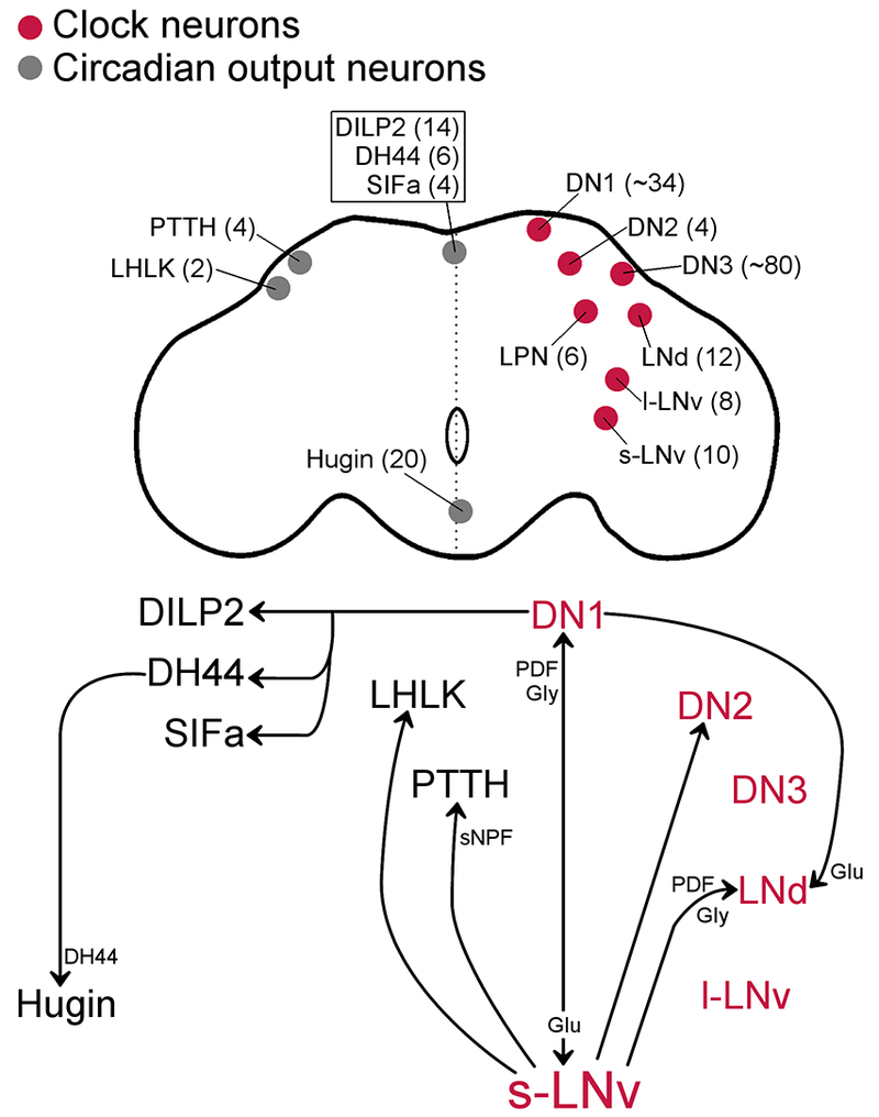
Top. Schematic representation of a fly brain with neuroanatomical locations of clock neurons (red, right hemisphere) and circadian output neurons (gray, left hemisphere or midline). Bilaterally represented neurons are labeled in only one of the hemispheres. Approximate total number of cells in the brain is indicated in parentheses. Bottom. Arrows represent the paths of communication between groups of circadian neurons. Circuits were mapped using neuronal activation and functional imaging and/or GRASP (GFP reconstitution across synaptic partners) methods. The neuropeptide/neurotransmitters that signal in the circuits were genetically identified by removing the peptide or neurotransmitter transporter in the presynaptic neuron and removing the receptor in the postsynaptic neuron. PDF mediates s-LNv communication to LNd, DN1, and LHLK (indirectly) (Leucokinin+ lateral horn). Glycine (Gly) also signals from s-LNv to DN1 and LNd. Short neuropeptide F (sNPF) signals in the s-LNv to PTTH circuit. Glutamate (Glu) signals from the DN1 to s-LNv and LNd. The molecules that signal between DN1 and PI neurons (DH44/SIFa/Dilp2) are unknown. DH44 neuropeptide signal from Dh44+ to hugin+ neurons.