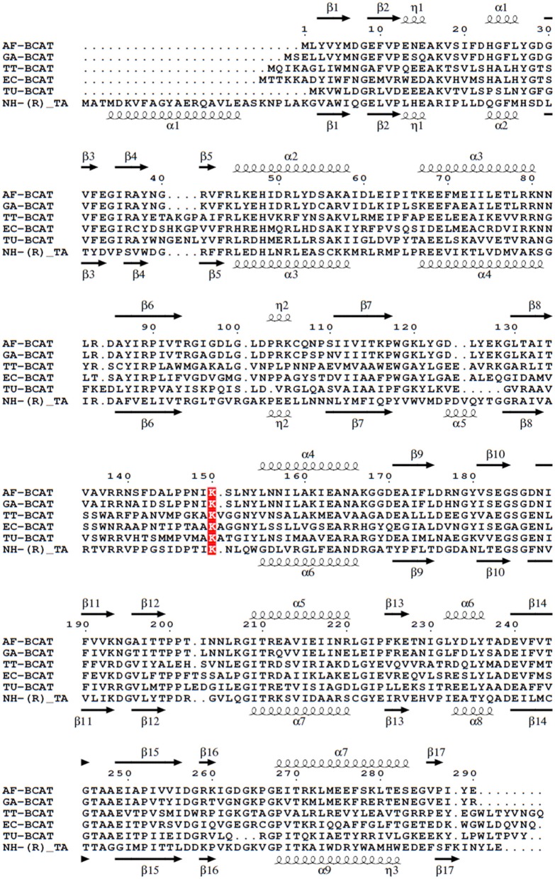
Figure 6.
Multiple sequence alignment of different BCATs, A. flugidus, G. acetivorans, T. thermophilus, E.coli, T. uzoniensis, N. haematococca. Arrows indicate β-strands, and helical curves denote α-helices of the structure of AF0933 above and Nectria TAm below. The active site lysine is highlighted in red. The figure was prepared with ESPript3 (Robert and Gouet, 2014).

