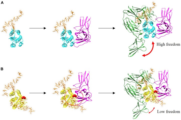
FIGURE 6.
Binding complexes formed by IFN-β-1a (A, cyan) and R27T (B, yellow) to binary and ternary assemblies with the ECD of IFNAR1 (green) and IFNAR2 (magenta) are presented with modeled structures. N-linked glycan at N80 and N25 residues (orange) are shown as sticks. A red sphere indicates residue R27T substituted in IFN-β-1a. Additional glycosylation of R27T induces the steric hindrance that affects its interaction with IFNAR2, and R27T likely has less conformational freedom than IFN-β-1a in the ternary complex.

