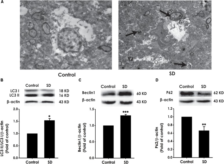
FIGURE 7.

Effects of SD on the autophagy in the hippocampus of rats. After 72 h exposure to SD, the rat’s hippocampus was separated. Autophagic vacuoles (A) was observed under transmission electron microscope. Arrows indicate autolysosome-like vesicles in the cytoplasm. LC3-II/LC3-I (B), Beclin-1 (C), and P62 (D) expressions in the hippocampus of rats were detected by Western blot using anti-LC3, -Beclin-1, and -P62 antibody, respectively. β-actin was used as loading control. Data are reported as the mean ± SEM (n = 3–5); ∗P < 0.05, ∗∗P < 0.0, ∗∗∗P < 0.001, vs. control group.
