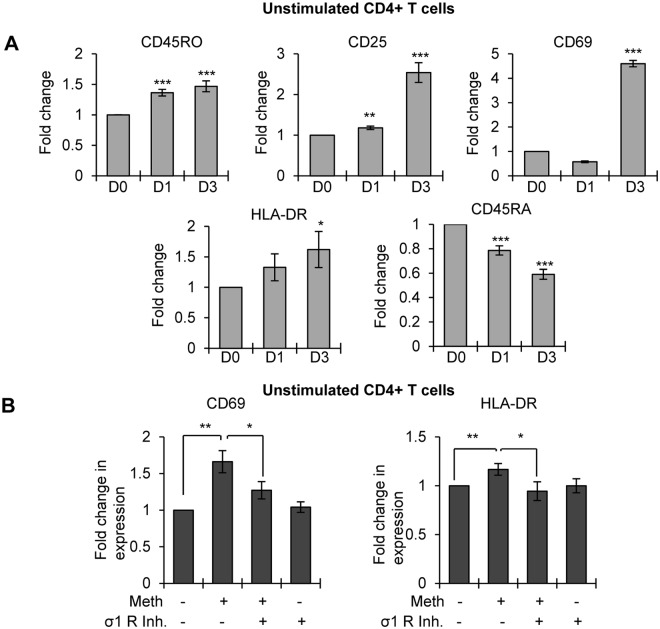
Figure 6.
Meth induced the expression of activation markers in unstimulated CD4+ T-cells: (A) Unstimulated CD4+ T-cells were treated with or without Meth (100 µM) for 3 days, cells were harvested on days 0, 1 and 3 (D0, D1 and D3), stained for the indicated T-cell activation markers, fixed and analyzed by flow cytometry. Bar diagrams showing the fold change in the expression of indicated activation markers in Meth treated unstimulated CD4+ T-cells. Fold change was calculated by normalizing the Meth treated cells to untreated cells (*p ≤ 0.05, **p ≤ 0.01, ***p ≤ 0.001). (B) Fold change in the expression of CD69 (left panel) and HLA-DR (right panel) in untreated or Meth treated unstimulated CD4+ T-cells in the presence or absence of sigma-1 receptor inhibitor (σ1 R inh.) after 3 days (*p ≤ 0.05, **p ≤ 0.01).

