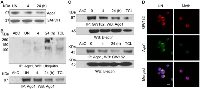
Figure 7.
Meth induced degradation of Ago1 and altered structural integrity of P-bodies: (A) CD4+ T-cells were untreated or treated with Meth (100 µM) for 0, 4 and 24 hours, lysed and Ago1 expression was analyzed by Western blotting. GAPDH used as a loading control. Full-length blots are presented in Supplementary Fig. S4 (B) CD4+ T-cell lysates in (A) were immunoprecipitated with Ago1 antibody and subjected to Western blot analysis using Ubiquitin antibody. Ago1 served as a loading control; AbC = Antibody control, TCL = Total cell lysate. Results are representative of 3 independent experiments. (C) CD4+ T-cell lysates in (A) were immunoprecipitated with GW182 antibody (upper panel) or Ago1 antibody (lower panel) and subjected to Western blot analysis using Ago1 (upper panel) or GW182 (lower panel) antibodies. B-Actin served as a loading control; AbC = Antibody control, TCL = Total cell lysate. Results are representative of 3 independent experiments. Full-length blots are presented in Supplementary Fig. S4 (D) Confocal images of GW182 and Ago1 interaction in CD4+ T-cells, untreated or treated with Meth (100 µM) for 24 hours. Scale bar = 10 µm. Results are representative of 3 independent experiments.

