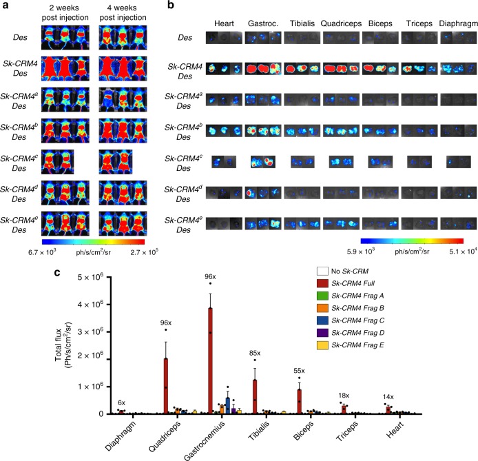
Fig. 3.
Screening and validation of Sk-CRM4 fragments. a In vivo bioluminescence imaging at week 2 and week 4 post vector injection and quantification of luciferase expression from the different individual organs of adult CB17/IcrTac/Prkdcscid mice injected intravenously with scAAV9-Des-Luc2 (control, no Sk-CRM), scAAV9-Sk-CRM4a-Des-Luc, scAAV9-Sk-CRM4b-Des-Luc, scAAV9-Sk-CRMc-Des-Luc, scAAV9-Sk-CRM4d-Des-Luc, and scAAV9-Sk-CRM4e-Des-Luc vectors at a dose of 1 × 10¹⁰ vg per mouse. The corresponding color scale from 6.70e + 03 (blue) ph/s/cm2/sr to 2.70e + 05 (red) ph/s/cm2/sr was shown. Photon emission was measured dynamically during 7 min in a supine position. b Ex vivo bioluminescence imaging of individual organs such as the heart and the different muscle groups harvested at 6 weeks post vector injection was represented on a color scale with luciferase intensities ranging from 5.92e + 03 (blue) ph/s/cm2/sr to 5.08e + 04 (red) ph/s/cm2/sr. The bioluminescence signal was quantified for 5 min. c Luciferase expression from the individual tissues was measured as total flux, expressed in photons/sec/cm2/sr. The fold-difference of the total flux was indicated for each Sk-CRM4 fragments relative to that of the control group containing the Des promoter without any Sk-CRM. The total flux data were displayed as mean + s.e.m. (n = 3)

