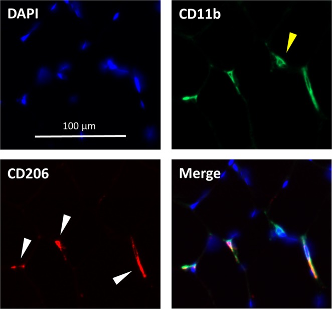
Figure 2.

Representative skeletal muscle macrophage immunohistochemistry. M2 macrophages were identified via co-stain for CD11b and CD206. Blue DAPI stain identifies nuclei. Green indicates CD11b, used as a pan-macrophage marker. Yellow carrot indicates a CD11b+/CD206- macrophage. Red indicates CD206, used to identify M2 macrophages. White carrots indicate cells that express both markers (CD11b+/CD206+). A merged image shows CD11b, CD206, and DAPI.
