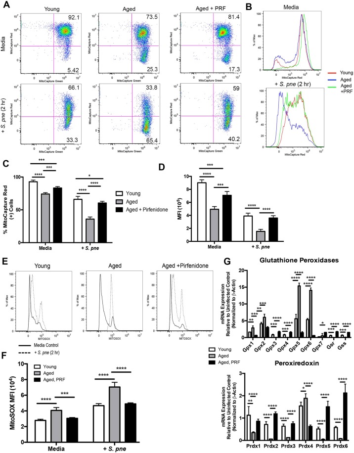
Figure 5.
Pirfenidone Treatment Improves Mitochondrial Membrane Potential and Decreases Superoxide Production in Aged Macrophages during S. pneumoniae Infection. Young macrophages were cultured with media alone for 24 hours prior to culture with S. pneumoniae (MOI = 25). Aged macrophages were cultured with media alone or media containing pirfenidone (500 μg/ml) for 24 hours prior to culture with S. pneumoniae (MOI = 25). (A,B) ΔΨm was examined in young, aged, and pirfenidone treated aged macrophages by assessing changes in MitoCapture red and green fluorescence in response to 2 hours of S. pneumoniae infection. (C) % of MitoCapture red positive cells and (D) mean fluorescence intensity (MFI) was assessed for each sample (t-test: *P < 0.05, **P < 0.01, ***P < 0.001, and ****P < 0.0001 and one-way ANOVA ****P < 0.0001). (E) Superoxide formation in young, aged, and pirfenidone treated aged macrophages at 2 hours post S. pneumoniae infection was measured using MitoSOX (solid line: media control, dashed line: S. pneumoniae infected). (F) Mean fluorescence intensity (MFI) was assessed for each sample (t-test: ***P < 0.001 and ****P < 0.0001 and one-way ANOVA ****P < 0.0001). (G) mRNA was isolated from untreated and pirfenidone treated aged macrophages at select time points of S. pneumoniae infection. Gene expression was analyzed by real time PCR using RT2 Profiler Assays (Mouse Oxidative Stress PAMM-065ZA). Specific mRNA expression of glutathione peroxidase (GPX) 1–7, glutathione synthetase (GSS), glutathione reductase (GSR), and peroxiredoxin (PRDX) 1–6 are illustrated (t-test: *P < 0.05, **P < 0.01, ***P < 0.001, and ****P < 0.0001 and one-way ANOVA ****P < 0.0001). Similar results were obtained from at least three independent experiments. Data are expressed as the mean ± SD.

