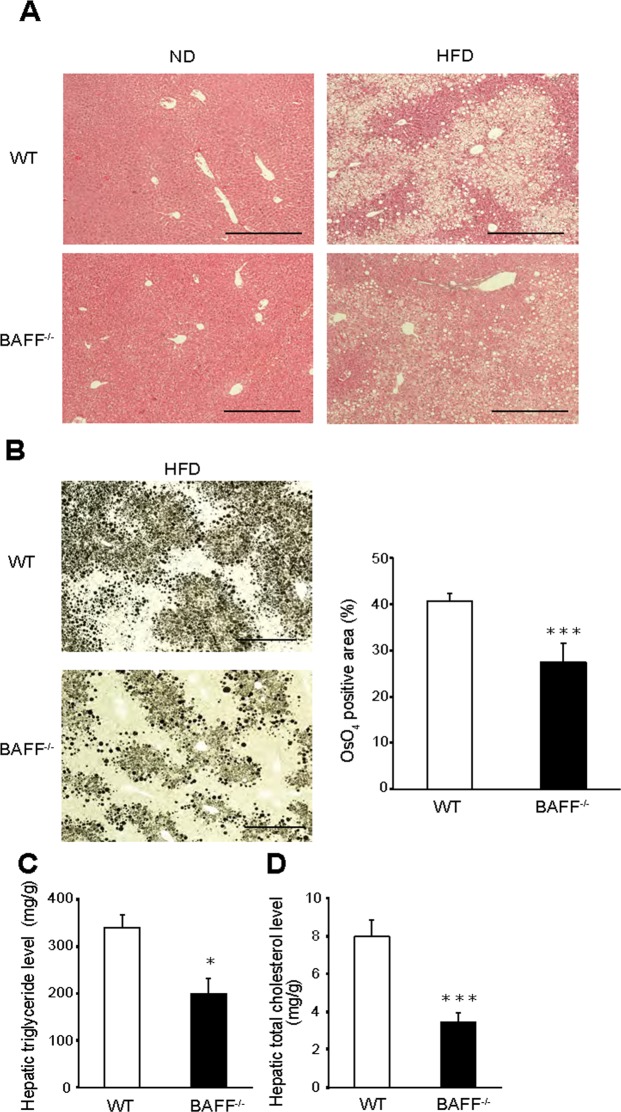
Figure 5.
Hepatic steatosis is ameliorated in HFD-fed BAFF−/− mice. (A) Representative H&E staining of livers from mice after 24 weeks of ND or HFD consumption (scale bar, 500 μm). (B) Representative OsO4 staining (left) and quantification of the OsO4-positive areas (right) of livers (scale bar, 500 μm; images from eight different fields; n = 8/group). (C) Hepatic TG and (D) total cholesterol levels (n = 5/group). For all bar plots, data are expressed as the mean ± SEM, and significance was determined by the Mann–Whitney U test. *P < 0.05 and ***P < 0.001. BAFF, B cell-activating factor; WT, wild-type; ND, normal diet; HFD, high-fat diet; TG, triglyceride.

