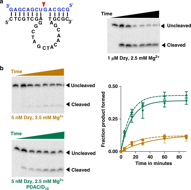
Fig. 7.
Coacervate-mediated stimulation of a DNAzyme. a Structure of the 10–23 DNAzyme. The enzyme strand is shown in black and the substrate is shown in blue. The red arrow indicates the cleavage site. Gel image shows efficient cleavage of the substrate (0.25 pM) by the enzyme (1 µM) in 25 mM Tris–HCl pH 8.0 containing 2.5 mM MgCl2 and 2.5 mM KCl. Time points were taken at 0, 2.5, 5, 15, 30, 60, and 90 min. b Reactions contained 5 nM of the enzyme strand and 0.25 pM substrate strand in 25 mM Tris–HCl pH 8.0 containing 2.5 mM MgCl2 and 2.5 mM KCl. Coacervates were formed by adding PDAC-53 and D10 at 10 mM total charge from each. Time points were taken at 0, 5, 15, 30, 60, 90, and 120 min. Fraction product formed were calculated from gels shown in b and Supplementary Figure 13 and data were fit to Eq. (1). Green solid and dashed lines indicate experiments performed in 2.5 mM Mg2+ or 5 mM Mg2+, respectively. All error bars represent S.E.M. (n = 3) from three independent experiments. Uncropped gel images are shown in Supplementary Figure 17b

