
Fig. 1. EZH2 interacts with USP44.
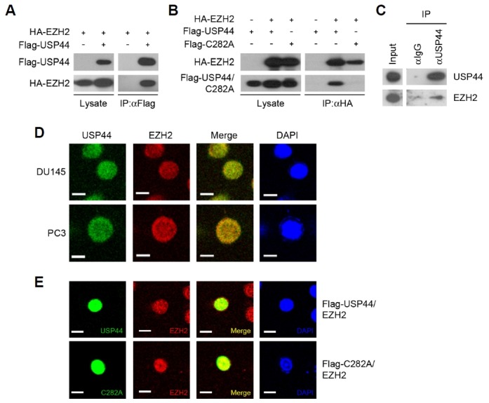
(A) HEK293T cells were transfected as indicated. Each cell lysate was immunoprecipitated with a Flag antibody followed by immunoblotting with Flag and HA antibodies. (B) HEK293T cells were transfected as indicated. Each cell lysate was immunoprecipitated with HA antibody followed by immunoblotting with Flag and HA antibodies. (C) Immunoprecipitation of USP44 from DU145 cell extract using an USP44 antibody followed by immunoblotting with USP44 and EZH2 antibodies. (D) Immunofluorescent staining of USP44 and EZH2 in DU145 and PC3 cells. USP44 was stained green and EZH2 was stained red. (E) DU145 cells were transfected with Flag-USP44 or Flag-USP44 C282A. Flag-USP44 or Flag-USP44 C282A was stained green and EZH2 was stained red. The blue signal represents nuclear DNA stained by DAPI. The bar indicates 10 μm.