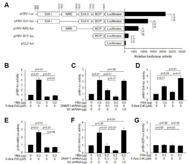
Fig. 3. HBx activates HBV core promoter via DNA methylation of NRE.

(A) Schematic representation of pHBV-luc and its derivatives. The basal luciferase activities of these constructs along with an empty vector pGL2-luc (Promega) were compared in HepG2 cells (n = 4). (B to G) Either an empty vector or HBx-expression plasmid was co-transfected with the indicated reporter construct into HepG2 cells for 48 h, followed by luciferase assay. The values indicate the relative luciferase activity compared to the basal level of the control (n = 4). (B, D, E, and G) Cells were either mock-treated or treated with 5 μM 5-Aza-2′dC. (C and F) The indicated amount of DNMT1 shRNA plasmid or SC shRNA plasmid was included in the transfection mixtures.
