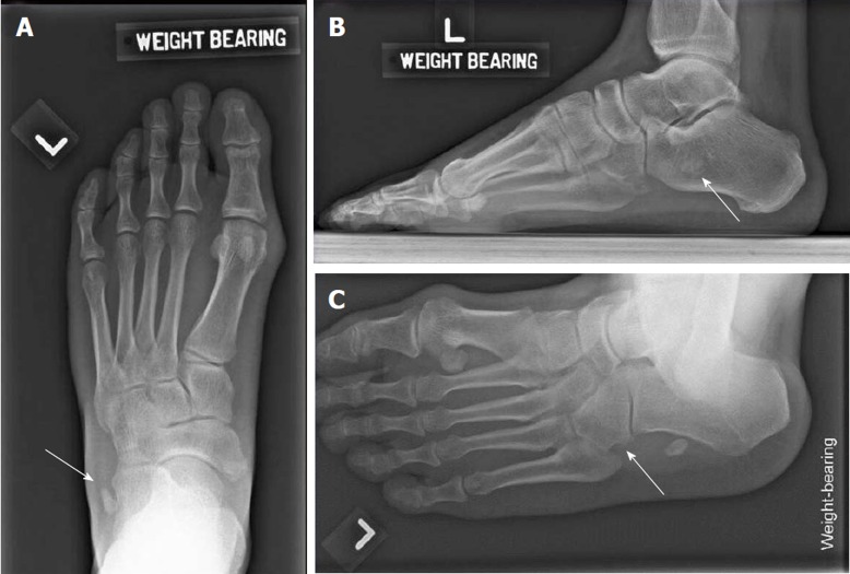
Figure 1.
Initial weight bearing radiographs of the injured foot. A: Weight-bearing dorsoplantar view of left foot showing well-defined bony fragment (white arrow) lateral to the anterior left calcaneum; B: Left lateral weight-bearing views shows a bony fragment at the level of the calcaneum; C: Left oblique view. White arrow shows the absence of the os peronuem at its usual anatomical. Instead, it was displaced proximally to the level of the anterior calcaneum process.

