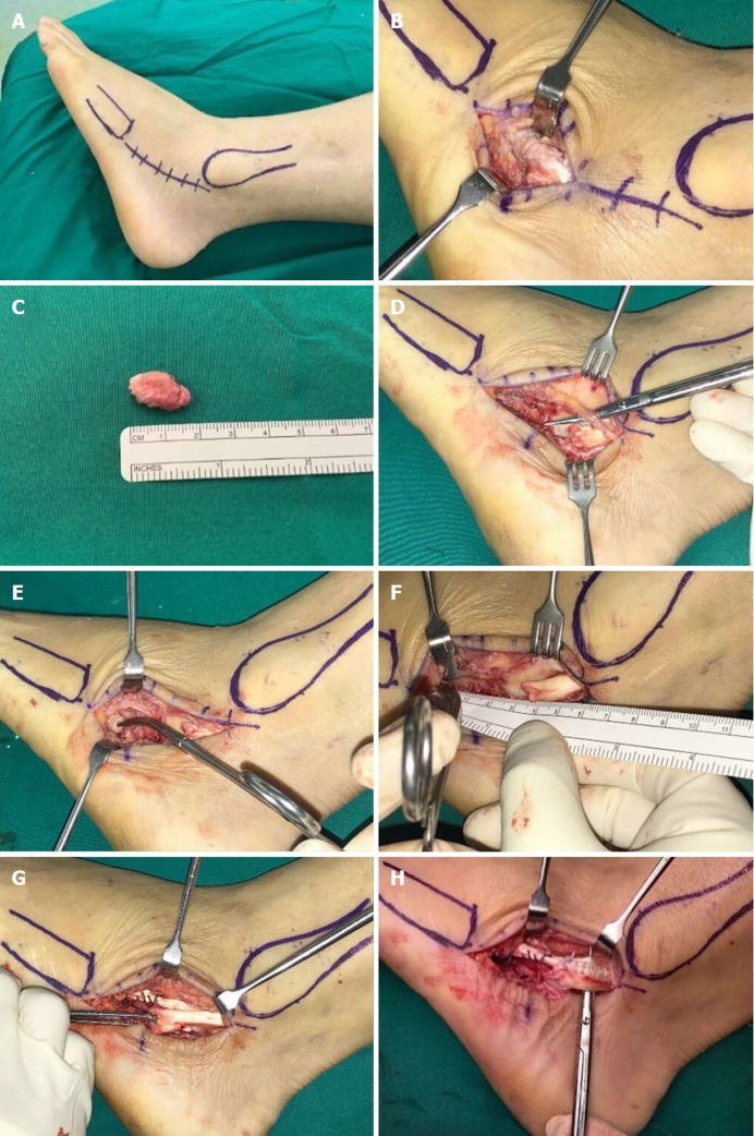
Figure 4.
Intra-operative images. A: Lateral position adopted with a thigh tourniquet applied; B: Incision was made from the left fibula tip to the base of the fifth metatarsal - centred around the os peroneum; C: The os peroneum was identified and excised and unhealthy tendon and devitalised synovium were debrided; D: The sural nerve was identified and protected during the surgery; E and F: Proximal and distal ends of the peroneus longus tendon (PLT) was mobilised and defect gap measured; G: The longitudinal split tear in the peroneus brevis was repaired; H: Side-to-side tenodesis of the PLT to the peroneus brevis tendon was performed.

