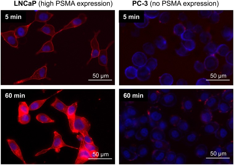
FIGURE 2.
Fluorescence microscopy (overlay) of internalization of [natLu]PSMA-I&F (100 nM) into LNCaP prostate carcinoma cells after 5 (top left) and 60 (bottom left) min at 37°C. Nonspecific background internalization was determined using PSMA-negative PC-3 cells (right). Red fluorescence = Cy5 filter (PSMA-I&F); blue fluorescence = DAPI filter (Hoechst 33342).

