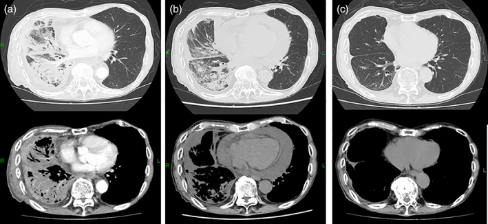
Figure 1.

Chest computed tomography (CT). (A) Prior to treatment with pembrolizumab. CT showed carcinomatous lymphangitis and pleural effusion. The primary site was not clear under cover to lymphangitis. (B) After six cycles. A massive pericardial effusion was observed. The other malignant lesions improved. (C) After 11 cycles. The patient showed a continuous good response.
