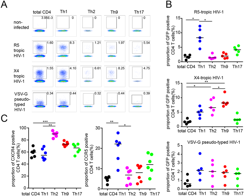
Figure 1: Increased susceptibility of Th9 and Th2 cells to X4-tropic HIV-1 in vitro.
(A-B): Proportions of GFP-positive CD4 T cells within indicated T helper cell subpopulations after in vitro infection with GFP-encoding X4-tropic, R5-tropic or VSV-G pseudotyped HIV-1. (A) shows representative flow cytometry dot plots from one study person, (B) demonstrates cumulative data from viral infections in PBMC from five HIV-1 negative study subjects. (C): Proportions of cells with CCR5 and CXCR4 surface expression in indicated CD4 T cell subpopulations. Data from six HIV-1 negative study subjects are shown. Data were tested for statistical significance using one-way ANOVA, followed by Dunn’s test for multiple comparison.

