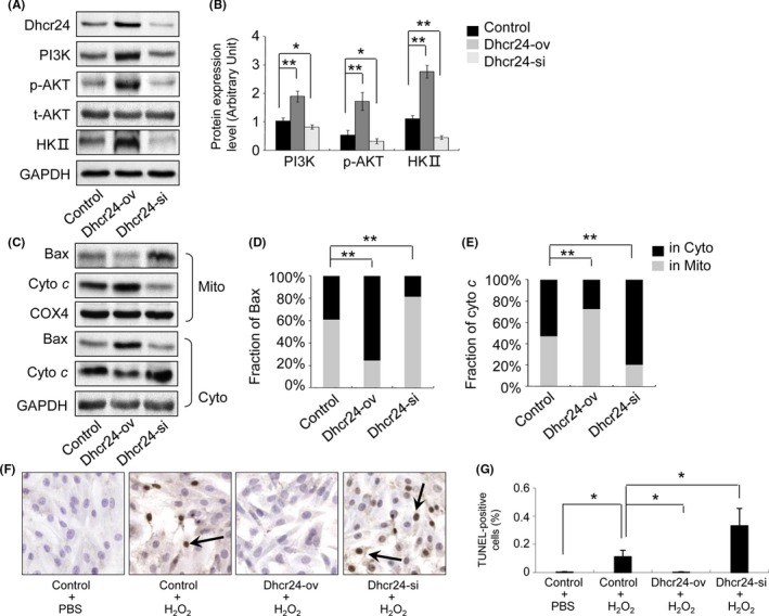
Figure 7.

The measurement of the signalling proteins of the PI3K pathway and apoptosis in H9c2 cells. The H9c2 cells, Dhcr24 over‐expressing (Dhcr24‐ov) cells and Dhcr24 knockdown (Dhcr24‐kd) cells were sampled, and the signalling proteins of the PI3K pathway and the mitochondrial pathway of apoptosis were detected by Western blot and TUNEL assay. (A), The levels of Dhcr24, PI3K, phosphorylated AKT and HKII were measured by Western blot. (B), Quantitative analysis using GAPDH for normalization (n = 3 independent experiments, *P < .05, **P < .01). (C), The fraction of Bax and cytochrome c in the cytoplasm and the mitochondria was detected by Western blot in the heart tissues. (D), (E), Quantitative analysis of the level of Bax and cytochrome c using COX4 and GAPDH for normalization, respectively, (n = 3 independent experiments, **P < .01). (F), Photomicrographs of the H9c2 cells and the Dhcr24‐ov and Dhcr24‐kd cells used for the TUNEL assay. The arrows indicate TUNEL‐positive cells. (G), Quantitative analysis of the apoptotic cells (magnification × 400, n = 3 independent experiments, *P < .05)
